LUNG ULTRASOUND LUS
Lung ultrasound can help diagnostic and monitoring patients with COVID-19, Cardiogenic pulmonary edema, Acute lung injury, Pneumothorax, Pneumonia, Interstitial lung disease, Pulmonary embolism, Chronic lung problem e.g. Interstitial lung disease, COPD.
Other important information follows:
Lung ultrasound is a relatively new method but in the next few years this technique is likely to become the standard of care in several Acute and Chronic medical problems not only of the lung but also heart, kidneys, and others.
A chest ultrasound is a noninvasive diagnostic exam that produces images from organs and structures within the chest.
Lung ultrasound has moved from its traditional assessment of pleural effusions and masses towards revolutionary approach of imaging the pulmonary parenchyma.
Portable devices and pocket-sized devices have also been proposed to perform lung ultrasound.
Another type of lung ultrasound is Doppler Ultrasound, sometimes called a duplex study, used to show the speed and direction of blood flow within the chest.
Lung Ultrasound may be safety used during pregnancy or in the pressure of allergies to contrast dye, because no radiation or contrast dyes are used
ADVANTAGES:
- All you need is a specialist cardiologist and the patient without large cumbersome machines
- Real Time dynamic imaging.
- No radiation
- Lung ultrasound can be very useful in neonates and children
- Anywhere and immediately
- Monitoring tool- Follow up
- Chest radiograph – chest CT-Lung ultrasound
- Meaningful evaluation in both inpatient and outpatient in both acute and chronic conditions
- Lung ultrasound can help diagnostic and monitoring patients with COVID-19, Cardiogenic pulmonary edema, Acute lung injury, Pneumothorax, Pneumonia, Interstitial lung disease, Pulmonary embolism, Chronic lung problem e.g. Interstitial lung disease, COPD.
Lung ultrasound is necessary in point of care ultrasound (POCUS), A method that can evaluate and diagnose an emergency or chronic medical problem in any patient.
It is necessary in various clinical settings from the emergency department to the intensive care unit for a patient with different underlying conditions but especially with heart, Lung, Kidney, and other Abdominal Problems.
In many circumstances Lung Ultrasound showing better sensitive and reliability that bedside chest X-Ray, especially in critically ill patients.
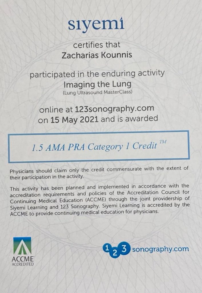
Dr. Zacharias Kounnis, has attended the course: Lung Ultrasound MasterClass “Imaging the Lung” and is constantly informed about the latest developments in this field.

Real Life Cases at our Cardiology Center
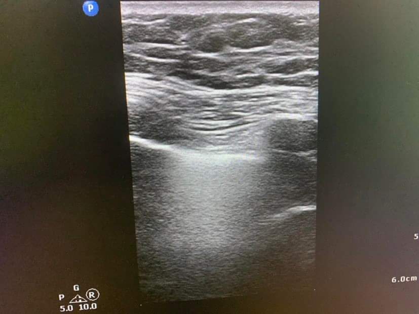
Case A: Normal Lung Ultrasound
Normal lung ultrasound. The pleura and the pulmonary parenchyma are clearly visible together with A Lines artefacts that characterize a normal lung.
Case B: A 54-year-old woman with Covid-19
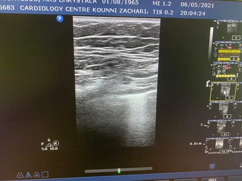
A 54-year-old woman who came to our cardiological center having already done three rapid tests negative two PCRs negatives and a chest x-ray on the same day and the report stated that everything was normal. Nevertheless, he continued to have shortness of breath and difficulty when breathing.
The following recordings were observed in the lung ultrasound compatible with Covid 19 despite the negative data it brought us. Finally, after a few hours, the patient’s oxygen dropped to 82 percent and he was hospitalized for 10 days in the intensive care unit suffering from COVID-19.


This incident shows the importance of lung ultrasound in combination with all the clinical laboratory findings in a person who may have symptoms from the respiratory system
Case C: A patient with Long Covid-19 Syndrome
A person who has already passed Covid-19 returned home and after almost a month could not return to his normal activity.
During his examination of the lung ultrasound, it revealed lesions due to Covid 19 that he had passed. We were dealing with a typical post Covid syndrome. Appropriate treatment was given and 10 days later the person had a great improvement in both his symptoms and the lung ultrasound.


